
Cochlear implants – the sound of innovation
Related topics
Health Innovation Health, Demographic Change and Wellbeing Austria Denmark Spain Switzerlanddate: 30/06/2015
Project: High-resolution image-based computationa...
acronym: HEAR-EU
See also: CORDIS
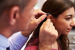
A cochlear implant is a small device, consisting of an implant placed under the patient’s skin and connected to an electrode array. This is inserted deep into the patient’s cochlea – the auditory part of the inner ear – during surgery, bypassing the non-functioning part of the cochlea.
An external audio processor picks up sound signals, which are then translated into electrical pulses and sent to the array, stimulating nerve fibres located in the cochlea. The hearing nerve receives these pulses and relays them to the brain, where they are perceived as sound, replacing the natural hearing system completely.
“Patients are all different, of course,” explains project coordinator Miguel A. Gonzalez Ballester of the Universitat Pompeu Fabra and the Catalan Institution for Research and Advanced Studies (ICREA) in Barcelona. “The outcomes of the surgery vary depending on how the operation is performed, which device is chosen, which electrode is implanted. So there is a need for more rationalised surgical intervention planning.”
One of the many challenges is to get highly detailed images of a specific patient’s hearing system, which is important because the bone and nerve structure will vary greatly. State-of-the-art imaging devices that could achieve a high enough resolution would expose the patient to too much radiation.
HEAR-EU got around this by studying the relevant structures in the ear and surrounding areas to build a high-resolution cochlear model which shows the range of variation in the population. This computational model, combined with a low-resolution image of the actual patient, is intended to help clinicians choose the best implant model and position for a patient’s anatomy.
A wealth of innovative results
However, to produce this extremely high resolution model, even for patients already implanted with the electrode, HEAR-EU first had to improve existing imaging technology, as the metal in implants will often create image noise, i.e. variation in brightness and colour in an image.
The resulting microCT device is expected to go to market very soon, says Gonzalez Ballester.
At the same time, the consortium – involving SMEs, industry and academia – is currently planning the exploitation of a first version of the surgical planning software, which will put to use the computational and statistical models developed during the project to help prepare and make recommendations for more tailor-made interventions. After assessing the number of potential users and the best exploitation model, the team plans to finalise its exploitation plan before the end of the project in August 2015.
In addition, HEAR-EU has come up with an improved cochlear implant design that will have a significant impact on the next generation of devices.
“As you can see, we are well on schedule on the technological front,” Gonzalez Ballester points out. “Some of the developments are already quite mature. For others, parts have already been validated, others still need to be integrated. The main task now for the remainder of the project is to run validations together with the clinicians. This will produce patient data and make sure that all of our software and computational models correlate well and lead to results that correspond to the clinical reality.”