Elektromagnetische Felder Aktualisierung 2009
3. Can mobile phones cause cancer?
- 3.1 Have studies on mobile phone users revealed an increased cancer risk?
- 3.2 Have studies on laboratory animals revealed an increased cancer risk?
- 3.3 Have studies on cell cultures revealed genetic effects?
- 3.4 Discussion on cancer
3.1 Have studies on mobile phone users revealed an increased cancer risk?
The SCENIHR opinion states:
3.3.2. Cancer
Studies on cancer in relation to mobile telephony have focused on intracranial tumours and acoustic neuromas because deposition of energy from RF fields from a mobile phone is mainly within a small area of the head near the handset. There are also some studies investigating the risk for other tumors in the head and neck region, notably tumors in the salivary glands. A small number of studies have investigated the association between exposure from RF fields from broadcast transmitters and tumour development. In animal studies, where sometimes whole body exposure is assessed, other forms of cancer have also been investigated. The in vitro studies considered aim to find out if biological effects relevant for carcinogenesis can occur at RF field levels that are typical for mobile telephony.
3.3.2.1. Epidemiology
What was already known on this subject?
In the previous opinion of 2007 a detailed discussion of epidemiological studies on mobile phone use in relation to brain tumour risk was presented. Altogether these studies provided evidence that mobile phone use for up to ten years is not associated with an increased risk of any type of brain tumours. With regard to a longer duration of use, uncertainties remained, as the number of such long-term mobile phone users was still small. Although none of the well-conducted studies indicated a substantial risk increase, they left the possibility open for a small-to-moderate risk increase among frequent mobile phone users, especially for glioma and acoustic neuroma.
What has been achieved since then?
Mobile phones and the risk of brain tumours
More data on mobile phones and brain tumours (including acoustic neuroma) became available from the Interphone study (Cardis et al. 2007), although the pooled analysis of data of the 13 countries involved has not yet been published. The Interphone study is a multinational case-control study coordinated by the International Agency for Research on Cancer (IARC). It is a population-based study with prospective ascertainment of incident cases and face-to-face interviews for exposure assessment. The new reports include the national studies from Norway (Klaeboe et al. 2007: glioma, meningioma, acoustic neuroma); France (Hours et al. 2007: glioma, meningioma, acoustic neuroma; article in French); Japan (Takebayashi et al. 2006: acoustic neuroma), Takebayashi et al. 2008 (glioma, meningioma); Germany (Schlehofer et al. 2007: acoustic neuroma); and two pooled analyses from the Nordic countries and the UK, one on glioma (Lahkola et al. 2007) and one on meningioma (Lahkola et al. 2008). Taken together, data based on about 60-70% of the total brain tumour cases of Interphone are published. As the remaining data were collected following the same study protocol, the already published majority of data defines limits of what is to be expected.
Most of the new Interphone reports were based on rather small sample sizes, particularly for acoustic neuroma (Takebayashi et al. 2006, Klaeboe et al. 2007, Hours et al. 2007, Schlehofer et al. 2007). For glioma and meningioma, the few new reports are consistent with the finding of no overall association derived from the previously published studies and hardly contributed to the state of knowledge about long-term mobile phone users (Klaeboe et al. 2007, Hours et al. 2007). The two pooled analyses by Lahkola et al. (2007, 2008) utilise larger numbers of subjects. However, as they combine the previously published data from Denmark, Sweden and the UK with new data from only Norway and Finland, the new insights are again limited. In the pooled analysis of glioma (Lahkola et al. 2007) including 1,521 cases, no increased relative risk was seen for long- term mobile phone users of ten years or more (odds ratio (OR) 0.95, 95% confidence interval (CI) 0.74-1.23). There were also no increased relative risk estimates for the highest categories of lifetime cumulative number of calls or lifetime cumulative duration of calls. In the meningioma pooled analysis (Lahkola et al. 2008) including 1,209 cases, most relative risk estimates were slightly decreased, e.g. for mobile phone users of ten years or more (OR=0.91, 95% CI: 0.67-1.25). The Japanese study was the first to go beyond analyses based on the years and the amount of mobile phone use (Takebayashi et al. 2008), by attempting to estimate the maximal specific absorption rate (SAR) value inside the tumour. No consistent pattern of relative risk estimates emerged from the use of various SAR values and both increased and decreased ORs were observed in the highest exposure categories.
Absorption of RF EMF from mobile phones is localised; therefore the preferred side of the head during mobile phone use becomes an important parameter of the exposure estimation. At the same time, this parameter is highly susceptible to reporting bias as cases know which side of their head is affected by the tumour, while controls do not know which side of their head will be relevant for analyses (in a matched study, it is the side of the head where the tumour occurred in their corresponding matched case). Therefore overreporting of the affected side of the head among cases may occur. This problem has already been identified in the very first case-control study on mobile phones and brain tumours using the approach of ipsi- and contralateral analyses (Hardell et al. 2002). An increased relative risk estimate for ipsilateral mobile phone use (preferred side of the head during mobile phone use corresponds to the side of the head where the tumour occurred) was compensated by a decreased relative risk estimate for contralateral mobile phone use (preferred side of the head during mobile phone use is opposite to the side of the head where the tumour occurred). This continued to be a problem in all subsequent case-control studies. Hence, the finding by Lahkola et al. (2007) for glioma risk among long-term mobile phone users of ten years or more received some attention when an observed increased OR for ipsilateral use was not compensated by an accordingly decreased OR for contralateral use (ORs of 1.39 versus 0.98, Table 1). An increased OR for ipsilateral use and an OR close to 1.0 for contralateral use would be expected under a hypothesized real effect. However, assuming causality, one would also expect that the effect of laterality becomes stronger with increasing exposure, i.e. the ratio of the two ORs for ipsilateral and contralateral use would be more or less close to 1.0 among short-term or occasional mobile phone users, but would then grow with increasing exposure. As displayed by the ratio between the ORs for ipsilateral and contralateral use in Table 1, this was not the case in the Lahkola et al. (2007) study, with laterality ratios being similar across exposure categories and being increased already among short-term users.
Hence, severe concern about reporting bias remains. Moreover, as the overall OR for long-term users was still below 1.0, the effect of an increased OR for ipsilateral use may be compensated by decreased ORs for cases with centrally located tumours or a missing value in the preferred side of use variable (data not shown separately). In conclusion, there is evidence that laterality analyses of retrospective studies are affected by reporting bias. It remains an open question, however, whether increased ORs observed for ipsilateral use in many studies are a mixture of a true effect and reporting bias or are due to such reporting bias in their entirety.
Two meta-analyses of case-control studies have been published on this topic since the last opinion statement (Hardell et al. 2008, Kan et al. 2008). No overall risk for brain tumors were found in the work by Kan et al. (2008), whereas both meta-analyses show an increased risk for brain tumors in long-term users (≥ 10 years). However, both studies are non-informative because of inappropriate exclusion criteria and combination of studies.
Table 1. Mobile phone use and the risk of brain tumours and parotid gland tumours
Validation studies conducted in the context of Interphone provided new information on the shortcomings of investigating this research question by means of an interview-based case-control study. Low response rates, particularly among controls, introduce bias if participation is related to mobile phone use (Cardis et al. 2007). This is a likely explanation for why many relative risk estimates in the Interphone study are actually below 1.0 (see for example Table 1). As ORs are particularly low in the subsets of shortest mobile phone use (Table 1), early symptoms before the diagnosis of the disease may impede regular mobile phone use among cases, which adds to the observation of a spurious protective effect. A comparison of self-reported mobile phone use and past traffic records from mobile phone operators confirmed previously observed general recall problems, but also indicated that there may be different patterns between cases and controls (Vrijheid et al. 2008). Although epidemiology is used to deal with imperfect measures, such bias-related uncertainty may seriously hamper straightforward conclusions.
Mobile phones and parotid gland tumours
In addition to the brain tumour data, the first two reports on parotid gland tumour risk have been published from the Interphone study. A pooled analysis of data from Denmark and Sweden showed no association with either short-term or long-term mobile phone use, but the study included only 60 cases of malignant parotid gland tumours (Lönn et al. 2006). Another 58 cases were accrued in the Interphone study in Israel (Sadetzki et al. 2008), showing similar results. The number of benign parotid gland tumours was larger in Israel (n=402) than in Denmark/Sweden (n=112) (Lönn et al. 2006). The Israeli study did not show an association with long-term use of mobile phones of ten years or more for benign and malignant parotid gland tumour combined (OR=0.86, 95% CI: 0.42-1.77). Although an effect of ipsilateral exposure was seen particularly in the subset of long-term users (Table 1), the relative risk estimate in this subset was decreased accordingly for contralateral use (Sadetzki et al. 2008).
Radio and television broadcast transmitters and childhood leukaemia
Prior to the previous opinion, there were only few studies on environmental radiofrequency electromagnetic fields (RF-EMF) exposure and the risk of cancer. As all these studies were ecological studies or cluster analyses, no conclusions could be drawn. Some studies, however, indicated an increased risk of leukaemia in children living close to strong radio or television broadcast transmitters (Ahlbom et al. 2004). Results from two case-control studies have recently become available. The first case-control study in South Korea involved 1,928 childhood leukaemia cases diagnosed between 1993 and 1999 and an equal number of hospital-based controls (Ha et al. 2007). RF-EMF exposure was calculated using a field prediction program and also the distance to one of 31 included amplitude-modulated (AM) radio transmitters was estimated. Although there was an excess of leukaemias in a 2 km radius of the transmitters (OR=2.15, 95% CI: 1.00-4.67), no association was seen between childhood leukaemia risk and the predicted field strengths (OR=0.83, 95% CI: 0.63-1.08 for the highest quartile of exposure); in the intermediate categories, relative risks were also decreased (revised main results table in the reply to a letter by Schüz et al. (2008)). The second case-control study was conducted in German municipalities surrounding 16 AM radio and 8 frequency-modulated (FM) radio and television broadcast transmitters (Merzenich et al. 2008). An exposure assessment for 1,959 childhood leukaemia cases diagnosed between 1984 and 2003 and 5,848 population-based controls was performed using field prediction programs. The main OR was 0.86 (95% CI: 0.67-1.11), comparing the upper ≥ 95% and lower < 90% quantile of the field distribution. No increased risk was seen for the first exposure decade alone, which was the time period without potential dilution from mobile telecommunication networks. The OR was 1.04 (95% CI: 0.65-1.67) among children living within 2 km of the nearest broadcast transmitter compared to those living at a distance of 10-15 km.
Source & ©: SCENIHR,
![]()
3.3.2 Cancer, p.16-19
3.2 Have studies on laboratory animals revealed an increased cancer risk?
The SCENIHR opinion states:
3.3.2.2. In vivo
What was already known on this subject?
The possible carcinogenicity of RF field exposure has been investigated in a number of experimental systems, with essentially negative results. The positive finding of increased lymphoma incidence in the lymphoma-prone transgenic Eµ-Pim1 mouse strain (Repacholi et al. 1997) is an interesting exception. The previous opinion of 2007 discussed a study (Utteridge et al. 2002) that failed to confirm the results of the Repacholi study, as well as several other studies that had evaluated carcinogenicity of RF fields in a variety of experimental models. Several studies had tested carcinogenicity of RF fields alone in normal or genetically predisposed animals, and several other studies had tested possible co-carcinogenicity together with known chemical or physical carcinogens. No statistically significant (p<0.05) increase of tumour incidence was found in any of the studies reviewed. Questions that remained were relevance of the experimental models to human carcinogenesis and the relatively low exposure levels used in most of the studies.
What has been achieved since then?
A number of lifetime and chronic exposure studies have been performed on laboratory animals.
The study reported by Oberto et al. (2007) was another replication and an extension of the Repacholi et al. (1997) study with Eµ-Pim1 transgenic mice exposed to a GSM-type signal. There were several methodological improvements compared to the original study by Repacholi et al. (1997) including use of several exposure levels (0.5, 1.4 or 4.0 W kg/kg), well-defined dosimetry and more uniform exposure (achieved by restraining the animals) and extensive histopathology of all animals. Compared to the sham-exposed controls, survival was reduced in the animals exposed to RF fields. The intergroup differences were statistically significant (p<0.05) in the male animals, but there was no trend with increasing exposure level (lowest survival at 0.5 W/kg). No increase in lymphoma incidence was observed in the RF exposed groups. Concerning other neoplastic findings, Harderian gland adenomas were increased in male mice, with a significant dose-related trend (p<0.01). However, this trend was not supported by the findings on female animals, i.e. no tumours were observed in the highest exposure groups. For the statistical analysis, the cage control and the sham-exposed control groups were combined, which is not a valid procedure given the differences in body weight development and tumour incidence between these groups (these differences are most likely related to restraint of the sham-exposed animals). However, based on the data reported in the paper, a different analysis strategy (comparison to the sham- exposed group only) would not essentially change the conclusion that there was no effect of RF electromagnetic fields on tumours at any site. The reduced survival in the exposed animals is not thoroughly discussed by the authors; this finding remains unexplained and difficult to interpret without detailed information about the causes of death.
In another study with lymphoma-prone animals (Sommer et al. 2007), unrestrained AKR/J mice, 160 animals per group, were chronically sham-exposed or exposed to a generic UMTS test signal for 24 h/day, 7 days/week at a SAR of 0.4 W kg/kg. No effect from exposure to RF electromagnetic fields was seen on lymphoma incidence, survival time or severity of the disease.
Two studies evaluated carcinogenicity of both a GSM signal at 902 MHz and a DCS signal at 1,747 MHz in conventional laboratory animals including B6C3F1 mice (Tillmann et al. 2007) and Wistar rats (Smith et al. 2007). Three exposure levels from 0.4 to 4 W/kg (and sham exposure) were used. The study on mice (Tillman et al. 2007) produced no evidence that RF field exposure increased the incidence or severity of neoplastic or non- neoplastic lesions, or resulted in any other adverse health effects. Interestingly however, the incidence of liver adenomas in males decreased with increasing exposure level, with a statistically significant (p<0.05) difference between the highest exposure and the sham exposed group. However, comparison with published tumour rates in untreated mice revealed that the observed tumour rates were within the range of historical control data. The study on rats (Smith et al. 2007) was a combined chronic toxicity and carcinogenicity study, and some of the animals (15 males and 15 females per group) were killed at 52 weeks from the start of the study. There were no significant differences in the incidence, multiplicity, latency or severity of neoplasms, or any other adverse responses to RF field exposure.
Saran et al. (2007) used Patched1 heterozygous knockout mice, an animal model in which exposure of newborn animals to ionizing radiation enhances development of brain tumours (medulloblastoma). Newborn Patched1 mice and their wild-type siblings were exposed to 900 MHz GSM-type radiation at 0.4 W/kg for 30 min twice a day for 5 days. No differences in survival were found between exposed and sham-exposed animals. Medulloblastomas (in 7 animals) and rhabdomyosarcomas (in 56 animals) were found in the Patched1 mice but not in the wild-type animals. The incidence of rhabdomyosarcoma was higher (68%, 36 animals) in the exposed group than in the sham-exposed group (51%, 20 animals), but this difference was not statistically significant (p>0.05). The incidences of medulloblastomas, other tumours or preneoplastic skin lesions did not differ between the exposed and sham-exposed groups.
Shirai et al. (2007) investigated possible promoting effect of 1.95 MHz RF fields (W- CDMA signal) on ethylnitrosourea (ENU)-induced brain tumours in Fischer 344 rats. The brain tumour incidences of both females and males tended to be higher in the two RF exposed groups (0.67 and 2 W/kg) than in the sham-exposed group, but no statistically significant (p<0.05) effects were reported. Moreover, an opposite trend (decreasing incidence with increasing exposure level) was observed in a previous similar study (Shirai et al. 2005), indicating that the trends observed are most likely incidental.
Hruby et al. (2008) treated 100 female Sprague-Dawley rats per group with 7,12- Dimethylbenz(a)anthracene (DMBA) to induce mammary tumours and then exposed the animals to 900 MHz GSM signals. The exposure groups included cage controls, sham- exposed controls and three exposure groups (0.4, 1.3 and 4.0 W/kg). The exposed and sham exposed animals were restrained during exposure. There were several statistically significant (p<0.05) differences between RF field-exposed groups and the sham-exposed group. All RF-exposed groups had significantly more palpable mammary gland tissue masses than the sham-exposed group, but there was no clear increase with increasing exposure level (no dose-response relationship). The incidence of malignant mammary tissue tumours was lowest in the sham-exposed group, and significantly increased in the high exposure group. However, the incidence of benign tumours was significantly lower in the three RF exposed groups than in the sham-exposed group. The number of animals with benign or malignant neoplasms was similar in the sham-exposed group and in the three RF-exposed groups. Given that the DMBA mammary tumour model is known to be prone to high variations in the results, the authors concluded that the differences between the groups were most likely incidental. Comparison with the results of the almost identical study of Yu et al. (2006) supports this conclusion: both studies reported similar development of mammary tumours in three groups, but lower rate of development (seen in the appearance of palpable tumours and/or reduced malignancy) in one group. Hruby et al. (2008) found the lowest rate of development in the sham- exposed group, while Yu et al. (2006) found it in the 0.44 W kg-1 group. Both studies consistently reported highest incidence of tumours in the cage control group, which is most likely related to the different handling of the cage control animals (different stress level, differences in food intake).
Source & ©: SCENIHR,
![]()
3.3.2 Cancer, p.19-21
3.3 Have studies on cell cultures revealed genetic effects?
The SCENIHR opinion states:
3.3.2.3. In vitro
What was already known on this subject?
In the previous opinion of 2007 a detailed description of in vitro studies was presented and discussed. Most of the studies did not provide evidence for any effect of RF field exposure at non-thermal intensity levels on cellular systems.
What has been achieved since then?
Many in vitro studies have been performed over the last two years. Both the genotoxic and non-genotoxic cancer-relevant effects are reviewed below.
Genotoxic effects
To test for genotoxicity, several techniques are available as no single assay is capable of detecting all genotoxic effects. These techniques include inter alia the micronucleus test (MN), DNA strand break test (comet assay), as well as tests for chromosomal aberration (CA) and sister chromatide exchange (SCE). The CA assay detects clastogenic (chromosomal breakage) or aneugenic (whole chromosome loss or gain) effects by direct examination of chromosomes in metaphase cells. The MN assay shows micronuclei containing nuclear DNA from chromosome fragments or whole chromosomes which were not incorporated into daughter nuclei at anaphase of mitosis. The assay detects the DNA damage in interphase cells. The SCE assay assesses DNA breakage and repair. SCEs are reciprocal exchanges of DNA segments between sister chromatids during S-phase. The molecular basis is unknown and SCE induction does not necessarily indicate mutagenicity. The comet assay or single cell gel electrophoresis (SCGE) quantify and analyse DNA damage in individual interphase cells, and can detect double strand breaks, single strand breaks, alkali labile sites, oxidative base damage, and DNA cross-linking with DNA or protein. It is also used to monitor DNA repair.
Several genotoxicity studies have been performed, with different outcomes. Using the MN test and the alkaline SCGE/comet assay no effects were detected in human lymphocytes, when exposure was administered at different stages of the cell cycle (24 to 68 h using 1950 MHz, 6 min RF on, 2 h RF off, 2.2 W/kg) (Zeni et al. 2008). Also, no increase in DNA strand breaks was found in trophoblast cells (Valbonesi et al. 2008). Schwarz et al. (2008) used the alkaline comet and the MN assays to study genotoxic effects of UMTS exposure (24 h, SAR 0.05 and 0.1 W/kg) on human fibroblasts and lymphocytes. They found that the exposure increased DNA damage at both SAR values according to these assays in fibroblasts, but not in lymphocytes. However, the scientific validity of this study is unclear, making any interpretation of the study difficult at this point.
Kim et al. (Kim JY et al. 2008) investigated mammalian cells exposed to 835-MHz RF fields (4 W/kg) alone, and in combination with a clastogen (ethylmethanesulfonate (EMS)). Genotoxicity was studied using the alkaline comet assay, and also CA. The combined exposure to RF fields and EMS revealed no significant (p>0.05) effects compared to EMS alone. However, the applied RF field had a potentiating effect in the comet assay, when administered in combination with another clastogen (cyclophosphamide or 4-nitroquinoline 1-oxide).
Another study investigating aneuploidy found changes in the number of certain chromosomes. Increased levels of numerical chromosomal aberrations (missing or extra chromosomes) were identified in human peripheral blood lymphocytes in vitro after 72 h RF field exposure (800 MHz, continuous wave, 2.9 and 4.1 W/kg). An induced aneuploidy of chromosomes 1, 10, 11 and 17 was determined. The increased levels of aneuploidy observed depended on the chromosome studied and on the level of exposure. In chromosomes 1 and 10, an increased aneuploidy was detected at the higher SAR value, while for chromosomes 11 and 17, the increases were observed only for the lower SAR value (Mazor et al. 2008).
Increased DNA damage was shown after RF field exposure in human lens epithelial cells (hLECs) (Yao et al. 2008). This effect was blocked by superimposing electromagnetic noise (2 µT). Twenty-four hour intermittent exposure (1.8 GHz, 1, 2, 3 and 4 W/kg) was used, and the alkaline comet assay and microscope detection of the phosphorylated form of histone variant H2AX (gammaH2AX) foci (a novel and very sensitive method to detect DNA strand breaks) was applied. DNA damage was significantly increased (p<0.05) after 3 and 4 W/kg exposures.
Non-genotoxic effects
In vitro analyses can be performed on different cellular processes such as cell cycle, induction of cell death (apoptosis), as well as metabolic and molecular changes.
In addition to DNA damage, Yao et al. (2008) investigated the induction of apoptosis and the release of reactive oxygen species (ROS) after 24-hour intermittent exposure to 1.8 GHz (1, 2, 3 and 4 W/kg) in human lens epithelial cells (hLECs). Significantly (p<0.005) elevated intracellular ROS levels were detected at 3 W/kg and 4 W/kg. After exposure to 4 W/kg, cells also exhibited significant (p<0.005) G(0)/G(1) arrest. However, no detectable difference in apoptosis was identified. All the effects were blocked when the RF-EMF was superposed with 2 µT electromagnetic noise. The authors suggested that the increased ROS levels may be associated with the induced DNA damage. No increased apoptosis rate was detected in primary cultured neurons from cerebral cortices of embryonic Wistar rats, exposed to 900-MHz GSM RF fields for 24 h (average SAR: 0.25 W/kg), using different techniques (Joubert et al. 2007).
The formation of ROS has been studied in L929 cells after exposure to 900 MHz RF fields with and without co-exposure to 3-chloro-4-(dichloromethyl)-5-hydroxy-2(5H)-furanonen (MX). Both continuous-wave and GSM mobile phone signals were applied for 10 or 30 min at specific absorption rates of 0.3 and 1 W/kg. The study provided no indication that 900 MHz RF-field exposure, either alone or in combination with MX, induces the formation of ROS (Zeni et al. 2007).
Using SH-SY5Y neuroblastoma and L929 cells, cell proliferation, oxidative stress and apoptosis were investigated in the presence of menadione (inducing ROS) or tert- buthylhydroperoxidase (inducing lipid peroxidation) and 872 MHz RF field exposure (CW or 217 Hz modulated, 5 W/kg for 1 or 24 h) (Höytö et al. 2008a). The authors detected an increased lipid peroxidation in SH-SY5Y cells using tert-buthylhydroperoxidase and 217 Hz modulated signal, whereas L929 cells showed an increased caspase 3 activity after co-exposure to menadione and a 217 Hz modulated signal. The other endpoints investigated (proliferation, viability, DNA fragmentation, glutathione levels) were not affected.
Höytö et al. (2007b) investigated the ornithine decarboxylase (ODC) activity in murine L929 fibroblasts using 872 or 835 MHz RF fields (CW or 50 Hz modulated) at 2.5 or 6.0 W/kg for 2, 8, or 24 h. The study was planned to replicate earlier studies reporting increased ODC activity in L929 cells but no effects were observed. In further studies by the same group, a lack of effects was confirmed in various cell culture conditions (Höytö et al. 2008b), and in several secondary cell lines (Höytö et al. 2007a) using 872 MHz CW or 217 Hz modulated fields at 1.5, 2.5, 5.0 or 6.0 W/kg. However, ODC activity in rat primary astrocytes was decreased consistently in all experiments performed at two exposure levels (1.5 and 6.0 W/kg), using GSM modulated or CW RF fields (Höytö et al. 2007a).
Exposure of the human trophoblast cell line HTR-8/SVneo to 1817 MHz sinusoidal waves (GSM-217 Hz; 1 h; SAR 2 W/kg) provided no evidence for increase of the HSP70- mediated stress response (Valbonesi et al. 2008). Another study showed that modulated 900 MHz RF fields (SAR 1 W/kg) lead to an anti-proliferative activity after 24 h in SH- SY5Y neuroblastoma cells, causing a G2-M arrest. In addition, markers of apoptosis were detected after a 24-h exposure, including a significant decrease (p<0.01) in mRNA levels of Bcl-2 and survival genes. On the molecular level it was also shown that RF field induced a transient increase in Egr-1 mRNA level paralleled with the activation of the MAPK pathway (Buttiglione et al. 2007).
It has been reported that mobile phone exposure (1.3 W/kg) alters the expression of a few proteins in human skin (Karinen et al. 2008); however the biological relevance of this finding is not clear. Zhao et al. (2007) studied the influence of 1.8 GHz RF fields on the gene expression of rat neurons. Among 1,200 candidate genes, 24 were up-regulated and 10 down-regulated after 24-h intermittent exposure at an average SAR of 2 W/kg. These genes are associated with multiple cellular functions (cytoskeleton, signal transduction pathway, metabolism, etc.).
Source & ©: SCENIHR,
![]()
3.3.2 Cancer, p.21-23
3.4 Discussion on cancer
The SCENIHR opinion states:
3.3.2.4. Discussion on cancer
Due to the introduction of mobile phone technology in the early 1980s and the beginning of its widespread use only in the mid 1990s, current epidemiological studies had difficulties in investigating brain tumour risk under the assumption of an induction period of about 10-20 years, because of the low number of users. The hypothesis of an induction period of more than 20 years could not be addressed in the current studies. In this context it needs to be noted that the digital mobile phone technology was only introduced in the early 1990s.
The evidence from epidemiological studies indicates that the use of mobile phones for less than ten years is not associated with an increased risk of developing a brain tumour. A major limitation of the current studies is that the diagnostic period of the cases ended in 2003 at the latest, hence, only a few long-term mobile phone users were included in those studies. This limitation together with uncertainties in reconstructing past exposures and difficulties in the ascertainment of representative study participants, circumvent firm conclusions related to long-term mobile phone use.
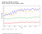
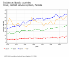
Altogether, the data collected until now provide no evidence of an increased brain tumour risk. This is consistent with the observation that no visible increases are seen in the age- specific incidence rates of tumours of the central nervous system in the Nordic countries over the last decade (Figure 1
, 2 ). As many more men (most of them between 30 and 60 years old) than women started to use mobile phones in the early days of the technology, an impact on incidence rates would be expected to appear first in men. A noticeable increase in the CNS tumour incidence rates from 1970 to the late 1980s, particularly in older men and women, is assumed to be an effect of improved diagnostic methods and appeared long before the widespread use of mobile phones. However, follow up of gender- and age-specific incidence rates remains important. However, despite new data, the existence of a small risk increase cannot be ruled out and therefore a similar level of uncertainty as in the previous SCENIHR report remains. This assessment may or may not change when the complete Interphone material is published. Prospective long-term follow up studies overcome both the limitations of retrospective exposure assessment and the latency problem and are recommended as a powerful long-term surveillance system for a variety of potential endpoints, including cancer, to fill current gaps in knowledge.
Recent well-conducted epidemiological studies provide evidence against an association between RF-EMF exposure from broadcast transmitters and the risk of childhood leukaemia. Although new exposure sources such as mobile phone base stations, cordless phone base stations or wireless networks are relatively recent, exposures from these sources are generally lower than the ones investigated in these studies on broadcast transmitters. Thus, there appears to be no immediate need for further studies related to these sources. However, no studies on mobile and cordless phone use among children and adolescents have been completed so far.
Seven recent studies with rodents have evaluated carcinogenicity of RF electromagnetic fields in vivo. Several different animal models were used including classical bioassays, studies using genetically predisposed animal models and co-carcinogenicity studies involving combined exposure to RF fields and known carcinogens. A few differences were reported for some endpoints, but no consistent dose-response pattern was observed, and the direction of the differences varied (increase or decrease in exposed animals), indicating that the few statistically significant differences are just statistical noise (false positive findings are unavoidable when many studies with multiple endpoints are conducted).
Overall, the results of the new studies are consistent with results from previous studies, and add to the evidence that the RF fields such as those emitted by mobile phones are not carcinogenic in laboratory rodents. Some of the new studies have also used exposure levels up to 4 W/kg which is high, compared to most previous studies. Thus, these studies provide additional evidence that carcinogenic effects are not likely even at SAR levels that clearly exceed human exposure from mobile phones.
Different biological endpoints have been investigated in vitro after RF field exposure using a variety of cell types and exposure conditions with diverse outcome. In the majority of studies no genotoxic effects were shown. A few studies suggest various biological effects (including genotoxic effects) from RF fields, alone or in combination with other factors, mostly at higher SAR values (above 2 W/kg). The biological relevance of these findings is however unclear. Inconsistent in vitro findings and a lack of dose response relationships render any mechanistic understanding of potential non-thermal interactions between RF and living systems difficult. For RF fields below the recommended limits (2 W/kg) for energy absorption due to mobile phones, in vitro studies have not identified reproducible effects by which carcinogenicity in living systems could be explained.
Source & ©: SCENIHR,
![]()
3.3.2, Cancer, p.23-26
GreenFacts asbl/vzw ist Inhaber des Urheberrechts der leserfreundlichen Drei-Stufen Struktur in welcher dieses SCENIHR Gutachten präsentiert ist.


